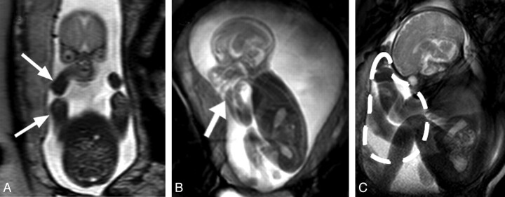
Fig 3.
Facilitation of flexed posture by the uterine wall. A, T2-weighted coronal section through a GA 22 weeks' fetus. The uterine wall is in close proximity to the fetus's lateral aspects (arrows). B, Still image from cine data of the same fetus. The fetal arm is flexed and located adjacent to the fetal head (thick arrow). C, Image of a GA 35 weeks' fetus demonstrates the redistribution of amniotic fluid volume near term, anterior to the thorax of the fetus (dashed line), due to its posture and the uterine limits; this collection of amniotic fluid may facilitate upper limb movements and trunk extensions.

