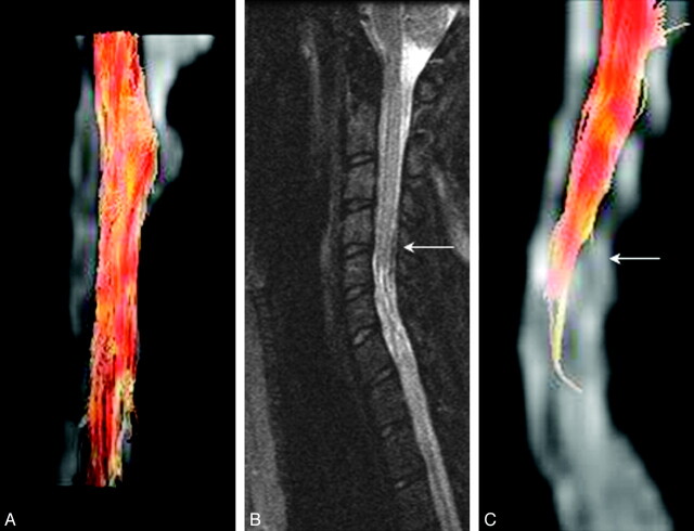
Fig 6.
A, MR tractography images of the cervical spinal cord of a child with IS derived from FA values in the white matter tracts, a measure of degree of myelination of the white matter tracts along the spinal cord. B, Conventional midline sagittal T2-weighted image of a child with SCI (complete injury, ASIA A). C, An MR tractography image based on the FACT algorithm of the cervical spinal cord of the child in B. This algorithm failed, however, to track the rest of the cervical cord well below the injury (arrow) level, even though the FA measurements for this subject 4 showed recovery of FA values as seen in Fig 2.

