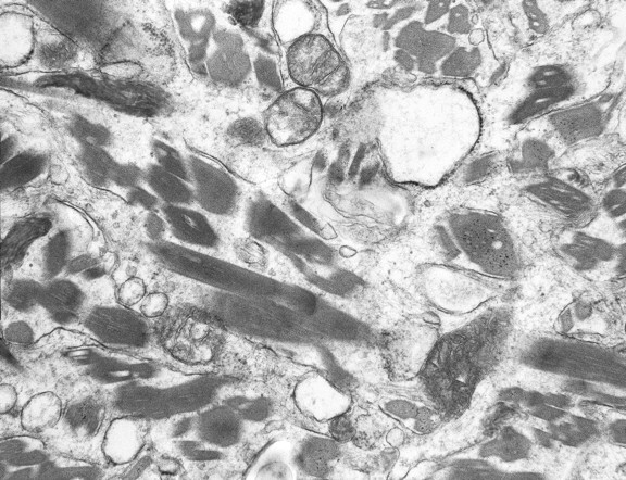
Fig 6.

Electron microscopy, ultrastructural photomicrograph shows abundant moderately osmophilic needlelike inclusions that appear geometrically irregular-to-rhomboid-shaped in cross-section within histiocyte cytoplasm. The inclusions show regular fine linear periodicity and are noted to occasionally be within phagolysosomes. No inclusions are seen within the mitochondria.
