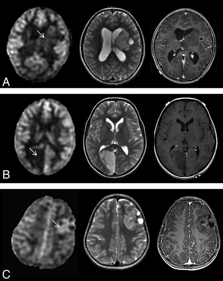
Fig 4.
ASL perfusion (left) of various low-grade tumors and correlative axial T2-weighted MR image (middle) and axial contrast-enhanced T1-weighted MR image (right). A, Low rTBF is seen within the pilocytic astrocytoma (arrow) with some tumor regions that show ASL signal similar to the contralateral gray matter in a 9-year-old boy. B, DNT shows low ASL signal (arrow) in a 9-year-old boy. C, Higher rTBF (arrow) compared with DNT (B) is seen in a 3-year-old girl with left frontal ganglioglioma.

