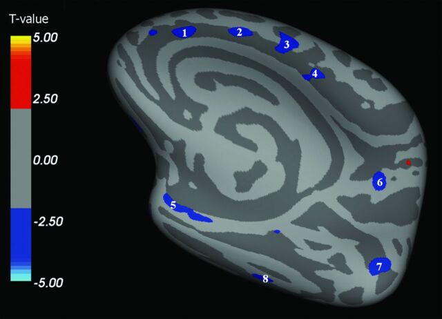
Fig 3.
Right hemisphere, medial surface. Areas showing a significant cortical thinning in patients with ALS (P < .05) are colored in blue. Areas 1–4 are localized in the frontal region. Areas 5 and 8 are localized in the temporal region. Areas 6 and 7 are localized in the parietooccipital region. CTh in area 4 (premotor cortex) correlated with disease duration (P = .007).

