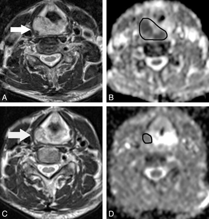
Fig 3.
A 68-year-old man with hypopharyngeal cancer (poorly differentiated squamous cell carcinoma). A, Pretreatment transverse T2-weighted MR image shows a primary hypopharyngeal cancer (arrow). B, The pretreatment ADC map derived from the DWI shows that the corresponding ADC value was 1.11 × 10⁻³ mm2/s for the manually placed region of interest covering the tumor. C, At 3 weeks after the start of treatment, the T2-weighted MR image shows a mass with marked regression (arrow). D, The ΔADCprimary at 3 weeks of treatment is 0.69.

