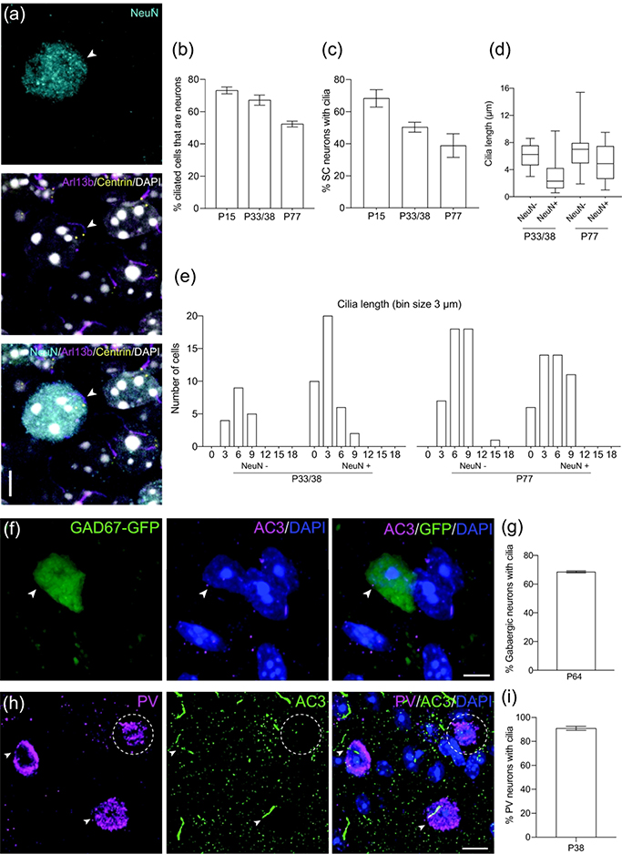
Figure 3.
Different types of neurons in the SC have cilia.
(a) Example of a P15 SC neuron (NeuN, cyan) extending a cilium (Arl13b, magenta). Centrioles labeled with GFP (yellow) and nuclei labeled with DAPI (white). Arrowhead indicates neuronal primary cilia.
(b) Quantification of ciliated SC cells that express the neuronal marker NeuN. Mean ± SEM. n = 91 P15, n = 374 P33/38, n = 218 P77 total cilia counted
(c) Quantification of SC neurons that have cilia. Mean ± SEM. n = 96 P15, n = 75 P33/38, n = 323 P77 total neurons counted
(d) Quantification of cilia length in non-neuronal cells and neurons in SC. n = 18 NeuN−, n = 38 NeuN+ P33/38 cilia; n = 44 NeuN−, n = 45 NeuN+ P77 total cilia measured
(e) Binned distribution of cilia length in non-neuronal cells and neurons in SC. The x-axis represents bin centers.
(f) Representative photomicrograph showing a cilium (AC3, magenta) extending from a P64 gabaergic SC neuron (GFP, green). Nuclei labeled with DAPI (blue). Arrowhead indicates co-expression of GFP and AC3.
(g) Percent of gabaergic neurons with cilia at P64. n = 162 total GAD67+ cells counted
(h) Representative photomicrograph showing cilia (AC3, green) extending from P38 parvalbumin (PV) neurons (magenta). Nuclei labeled with DAPI (blue). Arrowheads indicate co-expression of PV and AC3, and dotted ellipse indicates lack of overlap.
(i) Percent of parvalbumin (PV) neurons in the SC with cilia at P38. n = 108 total PV+ cells counted
Scale bar: (a,f): 5μm; (h): 10μm

