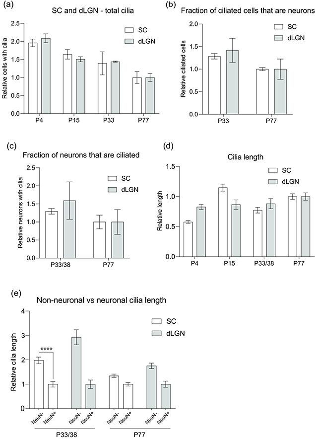
Figure 5.
Comparative analysis of the development of cilia in SC and dLGN.
(a-d) Normalized mean values (normalized to P77 of SC or dLGN, respectively) of total cilia in SC and dLGN (a), ciliated cells in dLGN and SC that are neurons (b), dLGN and SC neurons that extend cilia (c), and cilia length of dLGN and SC cells (d).
(e) Normalized mean values (normalized to NeuN+ at each age of SC and dLGN, respectively) of cilia length in non-neuronal cells and neurons; **** P<0.0001; Nested t-test (two-tailed).

