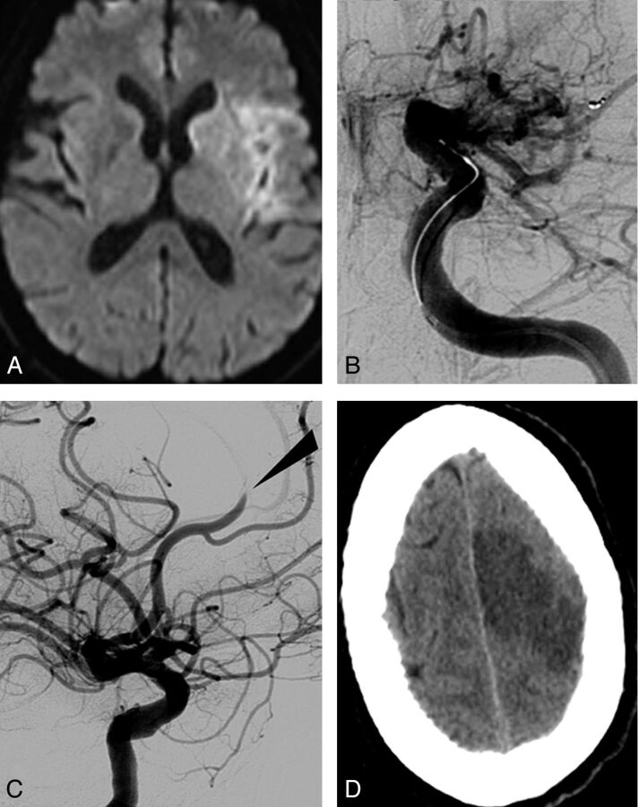
Fig 2.
A thromboembolic occlusion of the left anterior cerebral artery after clot removal from the ipsilateral MCA. A, Initial DWI showing ischemic lesion in the left MCA territory (DWI ASPECT score, 7). B, Left ICA Townes projection angiogram showing terminal occlusion. C, Lateral projection angiogram after removal of the device allowing flow restoration within the MCA but embolic migration within the left anterior cerebral artery (arrow). D, New ischemic lesion seen on CT after the procedure within the left anterior cerebral artery territory.

