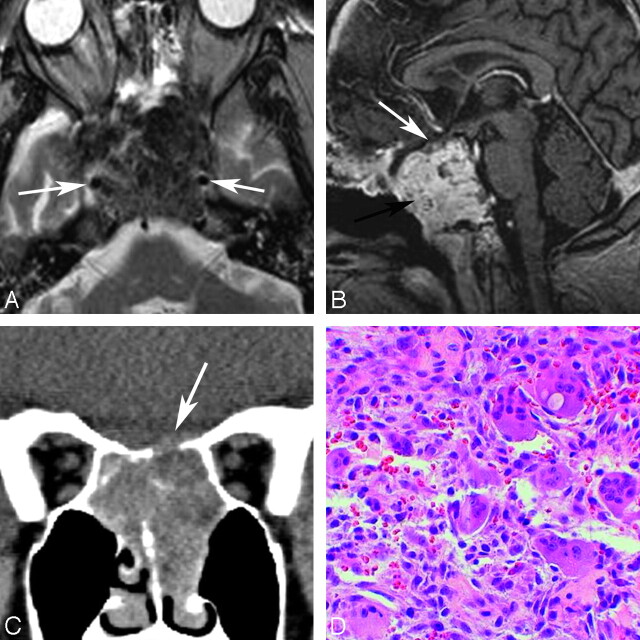
Fig 4.
Two patients with giant cell lesions of the sphenoid bone. A, An 18-year-old man with sphenoid giant cell tumor: axial T2WI shows relative hypointense areas within a lesion centered on the sphenoid bone and obliterating the sphenoid sinus cavity. These hypointense areas may represent areas of old blood products and fibrotic change. The carotids are displaced laterally (arrows). B, Gadolinium-enhanced sagittal T1WI shows the lesion avidly enhancing (black arrow). The pituitary gland and sella are displaced cephalad (white arrow). C, A 27-year-old woman with giant cell reparative granuloma. Coronal reformatted image in a soft-tissue window from a noncontrast CT scan shows a soft-tissue mass centered on the sphenoid sinus with dehiscence of the planum sphenoidale (arrow). D, HE stain on the photomicrograph shows giant cells with multiple nuclei, fibrous tissue, and blood products characteristic of this reactive lesion (original magnification ×40).

