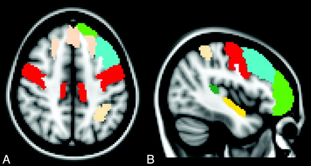
Fig 2.
Axial (A) and left parasagittal (B) normal T1-weighted templates show the cortical regions of interest in which a significant reduction of MTR was observed in patients with ALS with respect to healthy control subjects. These include the right and left precentral gyrus (red), right and left superior frontal gyrus (pink), frontal pole (green), planum temporale (yellow), left middle frontal gyrus (light blue), and left superior parietal gyrus (pink).

