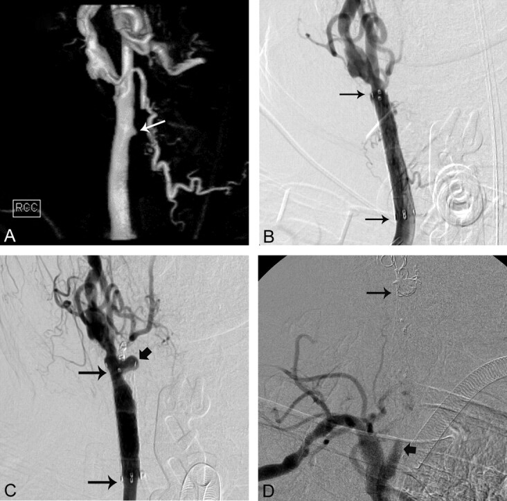
Fig 2.

A 65-year-old man with laryngeal cancer who presented with bright red blood from the tracheostomy site (On-line Table). A, 3D surface-rendering angiogram of the RCCA shows a small pseudoaneurysm (arrow). B, AP RCCA angiogram shows placement of a covered stent (long arrows) across the pseudoaneurysm. C, Three-month follow-up RCCA angiogram shows the development of a pseudoaneurysm (thick arrow) at the distal end of the covered stent (long arrows). D, After coil occlusion of the RCCA and RICA, a right brachiocephalic arteriogram shows occlusion of the RCCA (thick arrow) with coils within the RCCA (long arrow).
