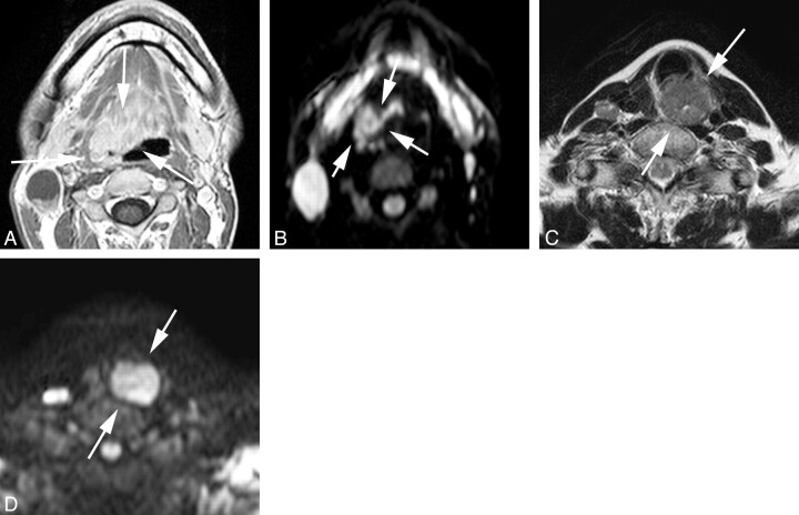
Fig 1.
Representative images of local control and failure cases obtained before treatment. A and B, Transverse gadolinium-enhanced T1-weighted and DWI (b = 1000 s/mm2) of a local-control case (oropharyngeal cancer, 30s, male, T4N2M0, high ADC before treatment = 0.63 × 10⁻³ mm2/s). C and D, T2-weighted and DWI (b = 1000 s/mm2) of a local-failure case (hypopharyngeal cancer, 60s, female, T4N2M0, high ADC before treatment = 0.99 × 10⁻³ mm2/s). The arrows indicate primary lesions.

