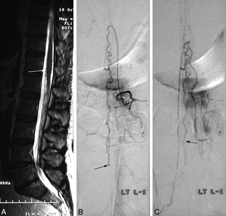
Fig 3.
Spinal AVF of the filum terminale (patient 1). A, Sagittal T2-weighted image shows high signal intensity in the conus with vascular signal-intensity voids (arrow) around the conus, raising the suspicion of a vascular spinal abnormality. B and C, Selective spinal angiograms show a feeding artery as a continuation of the anterior spinal artery, which is running downward to the site of the AVF on the filum terminale (L3 level, arrow in B) and the draining vein (arrow in C), which is running upward.

