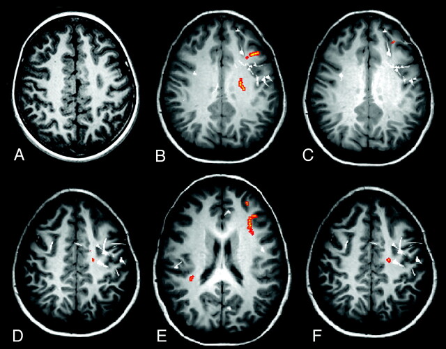
Fig 1.
Case 19. A, Axial volumetric T1-weighted image demonstrates thickening of the cortex in the left frontal gyrus (arrow), associated with blurring of gray-white matter, in keeping with FCD. Regions with abnormal FA (B), MD (C), λ1 (D), λ2 (E), and λ3 (orange areas) (F) and MEG dipoles (white) are overlaid onto volumetric T1-weighted image. The MEG dipole cluster, corresponding to the epileptogenic zone, is localized to the left frontal lobe and a few scattered dipoles in the right frontal lobe. There is lobar concordance of abnormal FA, MD, and eigenvalues with the MEG dipole cluster. There are bilateral abnormal λ2s, most of which are localized to the left frontal lobe. There is also overlap among the x-, y-, and z-axes distributions of abnormal FA, MD, and 3 eigenvalues with the MEG dipole cluster.

