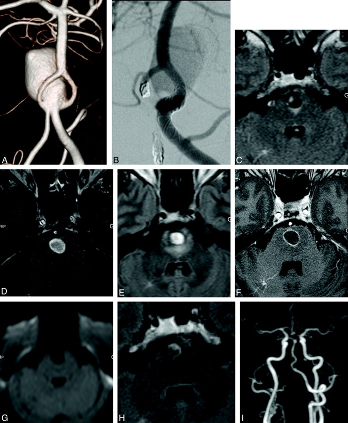
Fig 1.

A, The angiogram shows a giant aneurysm that developed at a fenestration of the vertebrobasilar junction. B, A study of the left vertebral artery just after deployment of the flow diverter shows stagnation of the contrast media and complete thrombosis 3 months postprocedure. C and D, In the same patient, the baseline MR image shows a small area with high signal intensity on FLAIR-weighted imaging and a circulating aneurysm on T1-weighted imaging without perianeurysmal enhancement. In the same patient, MR imaging repeated 10 days after Silk-treatment, while clinical symptoms are worsening, shows a wide area with high signal intensity on the FLAIR image (E), circumferential aneurysmal wall enhancement after contrast media (F), and no ischemic signs on trace diffusion-weighted imaging (G). In the same patient, 9 months after treatment, there is no longer peri-aneurysm edema on the FLAIR sequence (H), and the aneurysm is totally excluded from the circulation on MR angiography (I). Note total aneurysmal occlusion (I).
