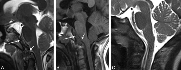
Fig 1.
Sagittal T2- (A) and T1-weighted (B) MR images of a 9-year-old girl (patient 4) who presented with craniocervical junction−related symptoms after an MVA show a REH (white arrow) with associated tectorial membrane disruption (short white arrow) and posterior dislocation of the dens. C, Sagittal T2-weighted MR image of an age-matched healthy control patient for comparison.

