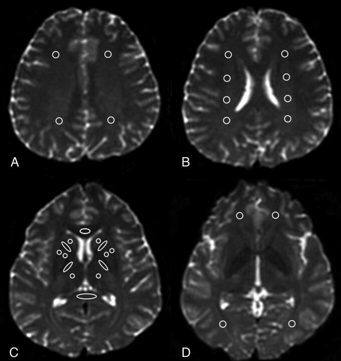
Fig 1.
Location of the ROIs in a healthy control. A, WM of the high frontal and parietal lobes located anterior and posterior to the central sulcus on the most caudal section on which they were visible and on the following cranial section. B, Periventricular WM was measured on the subsequent caudal section. C, ROIs at the genu and splenium of the corpus callosum were placed on 3 consecutive sections. The anterior and posterior internal capsules, caudate nucleus, globus pallidus, putamen, and thalamus were measured on 2 contiguous sections. D, Orbito-frontal WM was placed on the most caudal section of the lateral ventricles. Occipital-lobe ROIs were placed in the optic radiations of 2 contiguous sections, starting from the most caudal section on which the occipital horn of the lateral ventricle was imaged. Temporal lobe WM ROIs were placed lateral to the temporal horn of the lateral ventricle (not shown).

