
Fig 4.
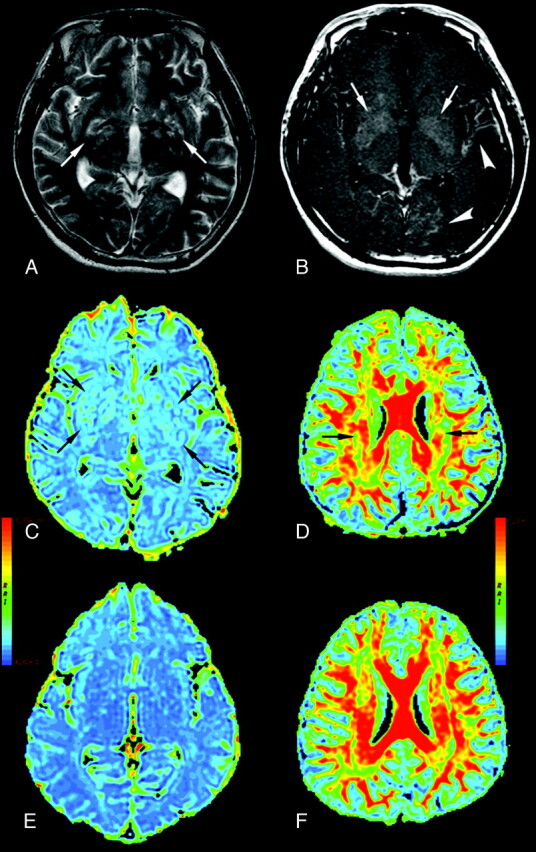
A 31-year-old man with CM (A–D) and a healthy control (E and F). A, Axial T2-weighted image. B, Axial T1-weighted image with contrast infusion displaying bilateral lentiform nucleus (arrows), left temporal and left occipital meningeal enhancement (arrowheads), C, ADC maps displaying bilateral dilated Virchow-Robin spaces (arrows) in the lentiform nucleus, and global cerebral ADC value increased. D, periventricular white matter FA value decreased (arrows).