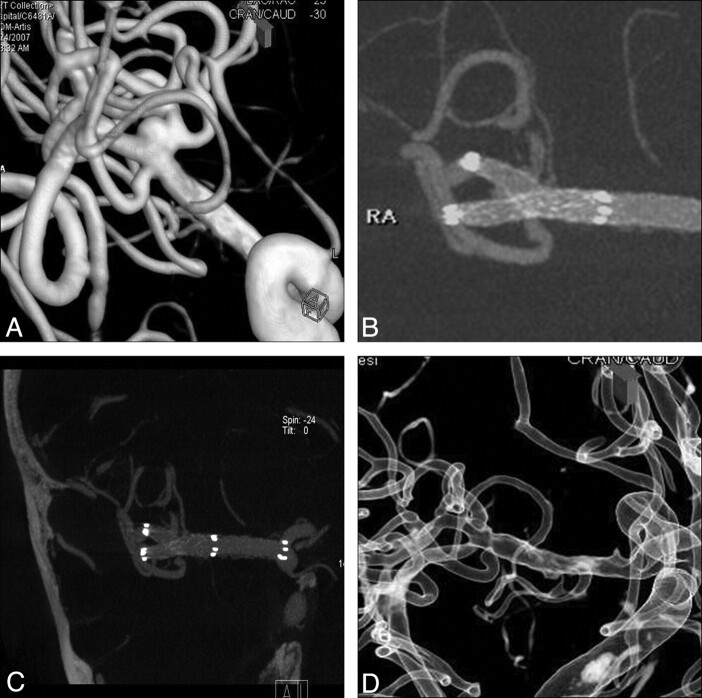
Fig 1.
Initial 3D reconstruction image of rotational angiogram (A) shows small wide-neck MCA trifurcation aneurysm. Posttreatment 3D reconstruction image of flat panel CTA (B) demonstrates the Y-configuration of the 2 stents placed within the MCA branches crossing the neck of the aneurysm, which is still filling in between. 3D reconstruction image of the flat panel CTA (C) obtained at 1-month control and 3D image of rotational angiogram at 1-year control (D) showing complete occlusion of the aneurysm and reconstruction of the trifurcation.

