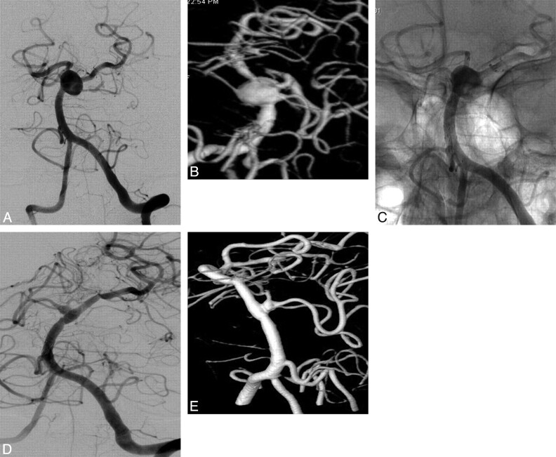
Fig 2.
Pretreatment left vertebral artery angiogram in oblique projection (A) and 3D reconstruction image of rotational angiogram (B) show the basilar tip aneurysm without a definable neck. Posttreatment angiogram (C), nonsubtracted view, shows the “Y”-configured stents extending into the both posterior cerebral arteries from the basilar trunk. The 2-year follow-up angiogram (D) and 3D reconstruction image of rotational angiogram (E) demonstrate completely remodeled basilar bifurcation with the aneurysm sac disappeared and occlusion of the right P1.

