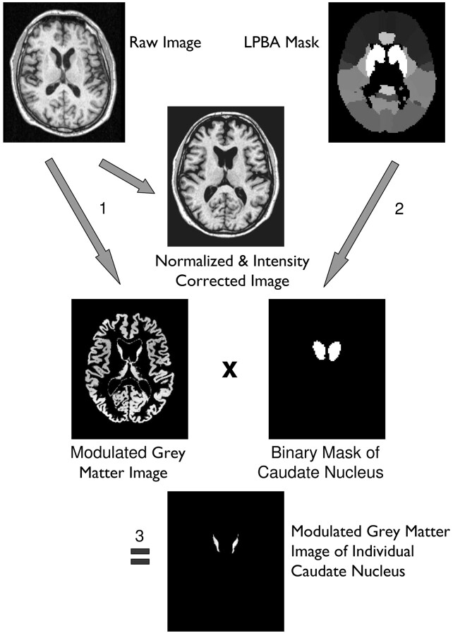
Fig 1.
Image processing and volumetry of the caudate nucleus: 1, Unified segmentation of SPM5 (ie, simultaneous normalization, segmentation, and intensity correction) is performed on a T1-weighted volume dataset. 2, A binary caudate mask is derived from the LONI Probabilistic Brain Atlas by setting all voxels of the maximum likelihood map belonging to the caudate nucleus to a value of 1, while all other voxels are set to zero. 3, The modulated gray matter image resulting from unified segmentation is multiplied by the caudate mask, resulting in a modulated image of the individual caudate nucleus.

