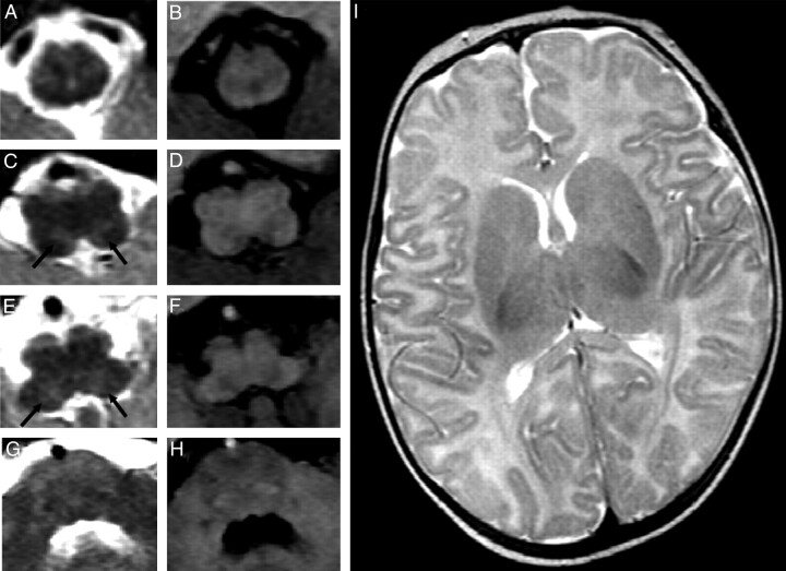
Fig 2.
MR images demonstrate the presence of tegmental lesions appearing on T2-weighted (A, C, E, G) and T1-weighted (B, D, F, H) images. Focal, bilateral, and symmetric lesions (arrows in C and E) show hyperintense signal intensity on T2-weighted images and faint hypointense signal intensity on T1-weighted weighted images. No signal-intensity or structural alterations are detected at the level of the cranial pons (G and H) and the supratentorial structures (I).

