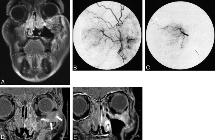
Fig 4.
Maxillary cancer with orbital invasion. A, This 59-year-old man was diagnosed with left maxillary cancer (T4N0M0) with orbital invasion. B and C, Before treatment, both an external carotid angiogram and a superselective internal maxillary angiogram reveal a tumor stain in the maxillary sinus. D and E, A subtraction image of the CT angiogram reveals the intraorbital component of the tumor as a hypoenhanced lesion (white arrows). After 4 sessions of intra-arterial infusion therapy, the intraorbital tumor has shrunk remarkably and is much more enhanced (E) than in the initial CT angiogram (D).

