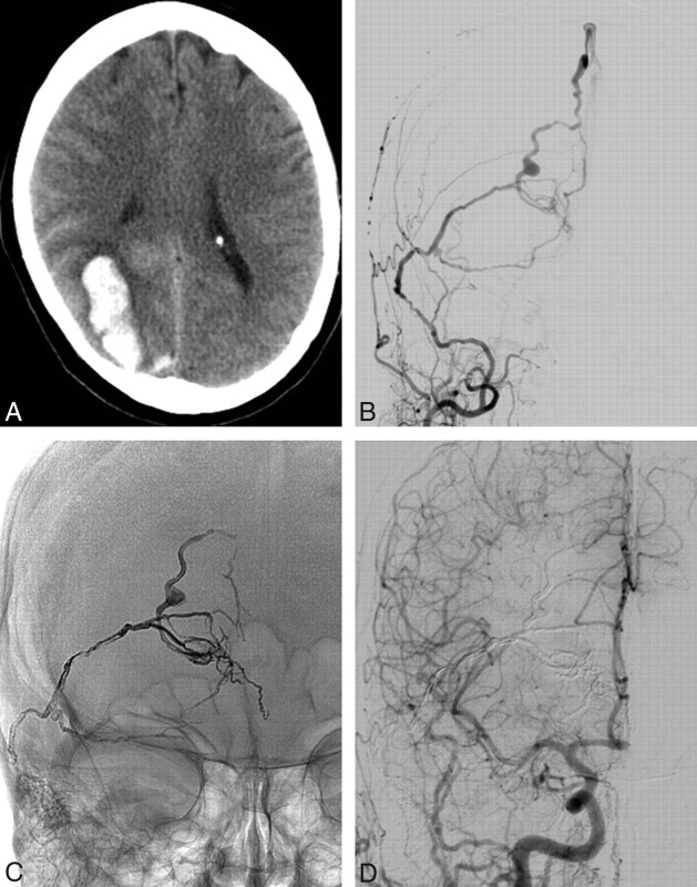
Fig 3.
A 59-year-old woman (patient 5) with a right occipital parenchymal hemorrhage from a DAVF. A, CT scan demonstrates a right parenchymal hematoma. B, AP view of a right external carotid angiogram shows a DAVF mainly supplied by the middle meningeal artery. Note the aneurysm on the occipital draining vein. C, Onyx cast after embolization through the middle meningeal artery. All dural supply and the draining veins together with the venous aneurysm are occluded. D, Follow-up carotid angiogram after 12 weeks confirms complete occlusion of the DAVF.

