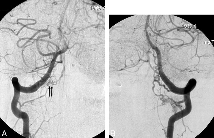
Fig 2.
Case 7. Coincidental bilateral spinal DAVS. A 39-year-old man presented with SAH. A, The anteroposterior view of the right VA angiogram shows a small dural shunt with the feeder from the right C1 dural artery (arrows). Venous drainage is rostral. B, The anteroposterior view of the left VA angiogram also reveals a separate dural shunt from the C2 dural artery, which also shows rostral venous drainage. Embolization was unsuccessful due to superselection failure; thus, surgical resection was performed for the respective lesions. The patient showed good recovery on 20-month clinical follow-up.

