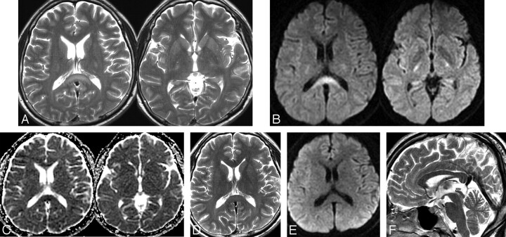
Fig 2.
Follow-up MR images obtained on days 17 (A–C) and 30 (D–F). A, On axial T2WI, hyperintense lesions are seen in the splenium and internal capsules. B and C, On axial DWI and an ADC map, the lesions appear hyperintense in DWI and hypointense on the ADC map in the splenium and internal capsule. D–F, On follow-up MR images obtained 3 months after onset, note the disappearance of all signal-intensity abnormalities on axial T2WI, DWI, and sagittal T2WI.

