
Fig. 3.
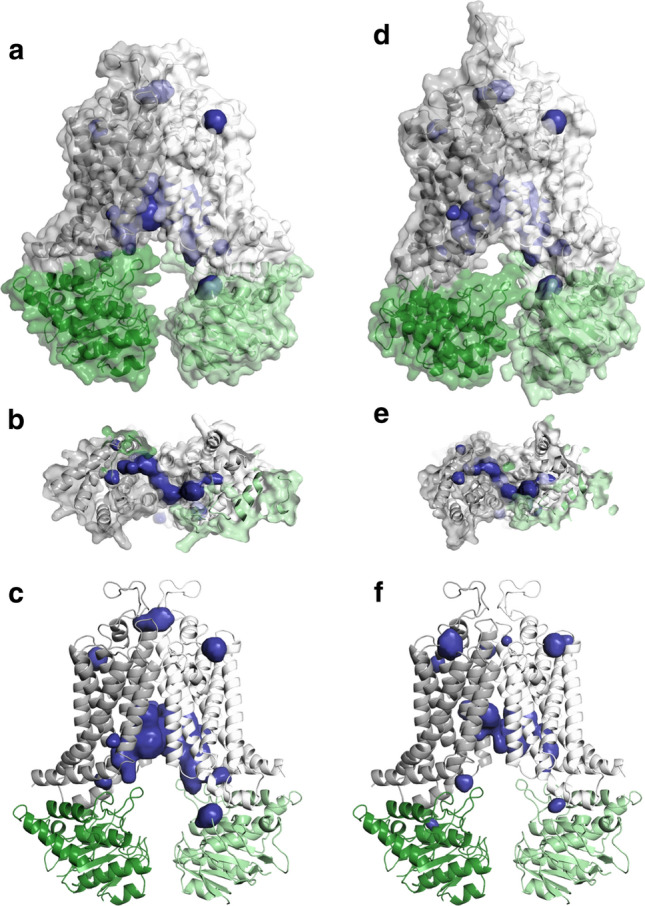
ABCG2 drug binding cavities in MD simulations correspond to pockets identified by in silico docking. a–c Binding pockets determined in an MD trajectory are shown in the context of the starting structure. d, e The closure of the NBD and TM helices limits access to the translocation pathway. f In some of the simulations, spaces forming a translocation pathway are less frequent and less continuous. Gray and white: transmembrane domains, green and light green: nucleotide-binding domains, blue: pockets