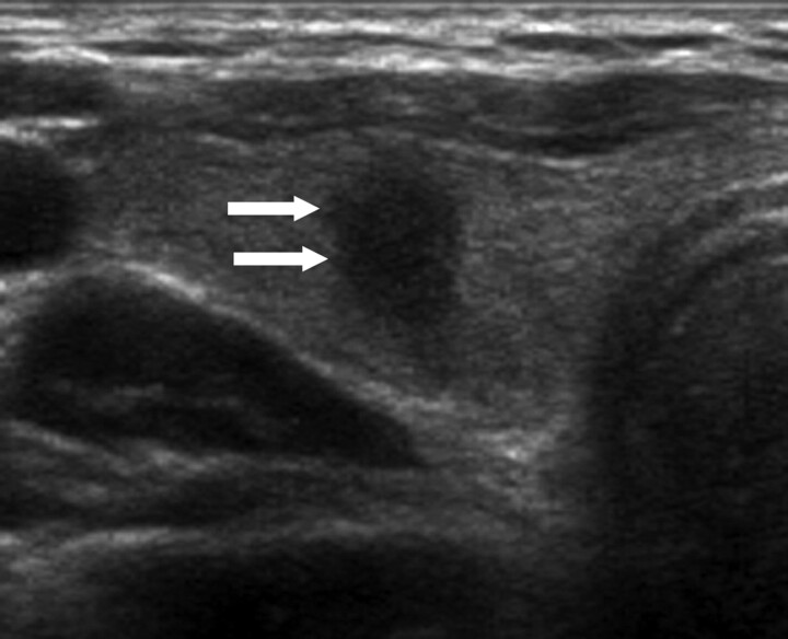
Fig 3.
A 42-year-old woman with papillary thyroid carcinoma. Transverse sonogram shows a 0.6-cm-long spiculated taller-than-wide hypoechoic nodule (arrows) within the right lobe of the thyroid gland, which is suspicious for malignancy by US criteria. Note there is neither a low-echoic halo nor an inner isoechoic rim at the periphery. Subsequent cytologic and histologic examination confirmed the diagnosis of papillary thyroid carcinoma.

