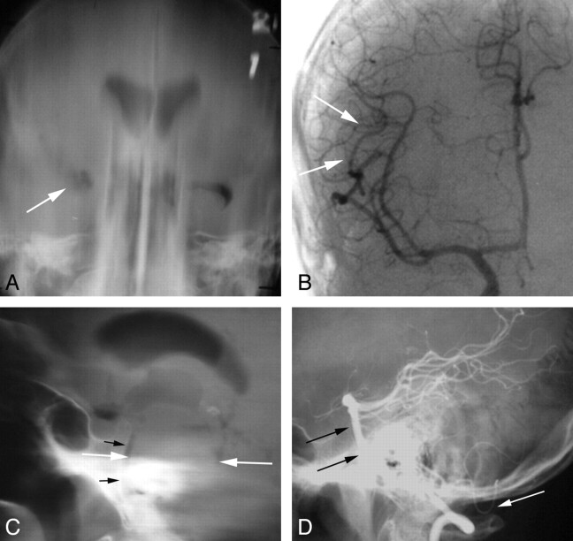
Fig 2.
Imaging of tumors with pneumoencephalography and angiography. A 48-year-old man with new-onset seizure. A, Frontal film from a pneumoencephalogram shows deformity and displacement of the right temporal horn (arrow), indicating a nonspecific mass in the anterior, basal, and lateral right temporal lobe. B, Frontal view from a right ICA angiogram shows minimal displacement of the right MCA branches in the Sylvian fissure (arrows). An infiltrating glioma was found at surgery. A 45-year-old man who presented with left-sided sensory changes of the face and body. C, Lateral film from a pneumoencephalogram shows marked enlargement of the pons (white arrows), which is nearly in contact with the clivus (black arrows). D, Lateral film from a vertebral angiogram shows anterior displacement of the basilar artery to the clivus (black arrows) and posterior inferior displacement of the posterior inferior cerebellar artery (white arrow). Findings are compatible with a brain stem lesion such as glioma, metastasis, or vascular malformation with hemorrhage. A glioma was found at surgery.

