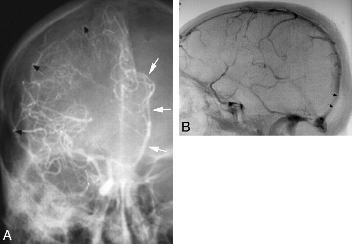
Fig 4.
Before CT and MR imaging, angiography was used to diagnose extra-axial hematomas in patients with acute head trauma. A, Frontal view from a right ICA angiogram shows displacement of right MCA branches away from the inner table of skull (black arrows) by a crescentic collection compatible with a SDH. There is also a distal-type midline shift with the anterior cerebral artery displaced across the midline (white arrows). B, Lateral angiographic image shows anterior displacement of distal superior sagittal sinus and torcular herophili (arrows), compatible with stripping of the dura from inner table of skull due to an EDH.

