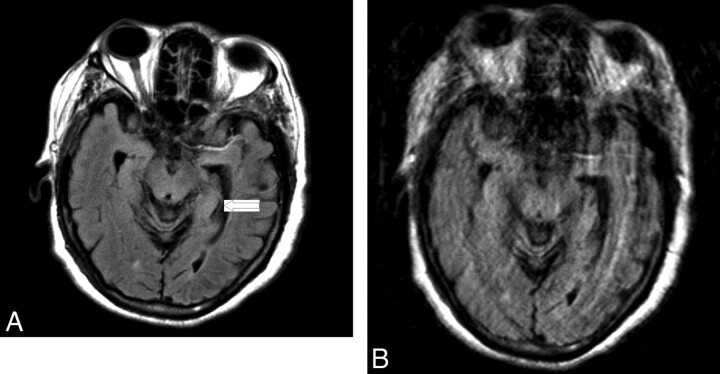
Fig 4.
Axial FLAIR images of patient who presented with an acute left middle cerebral artery distribution stroke obtained by using (A) BLADE and (B) rectilinear techniques. The partially radial acquisition image is free of motion artifact, whereas extensive motion artifact degrades quality of the rectilinear image. The small, subtle ischemic lesion was also better characterized from the partially radial acquisition image (arrow).

