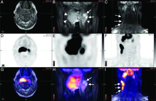
Fig 1.
A 44-year-old man with squamous cell carcinoma of the nasopharynx. Axial T1 (A) and coronal STIR (B and C) sequences, with corresponding PET (D–F) and the fusion of both (G–I), in a patient with nasopharyngeal carcinoma. The neoplastic tissue (arrowheads) is easily identifiable in STIR sequences (B) in which the cystic component can be visualized (arrows). The T1-weighted sequences allow a good assessment of the fat planes. Metastatic lymph nodes (dotted arrows) can be assessed morphologically (C) and functionally (I). Notice the concordance of the cystic areas of the lesion with an area of lower 18F-FDG uptake as expected.

