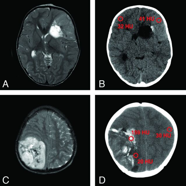
Fig 6.
T2WI of 2 EBLs with corresponding CT scans of patient 12 (A, B) and patient 19 (C, D). Note almost T2 isointense signal of the tumor in (A) with slight hyperattenuation in the corresponding CT image (B). This tumor is indicative of higher cellularity, compared with the EBL shown in (C) and (D). Calcifications such as in (D) can complicate the detection of components with high cellularity. Region of interest in red showing Hounsfield units of tumors, normal cortex, and calcifications (A, 1.5T Signa Excite, GE Healthcare; B, Sensation 16, Siemens; C, 0.5T NT Intera, Philips Healthcare, Best, the Netherlands; D, Lightspeed Plus, GE Healthcare).

