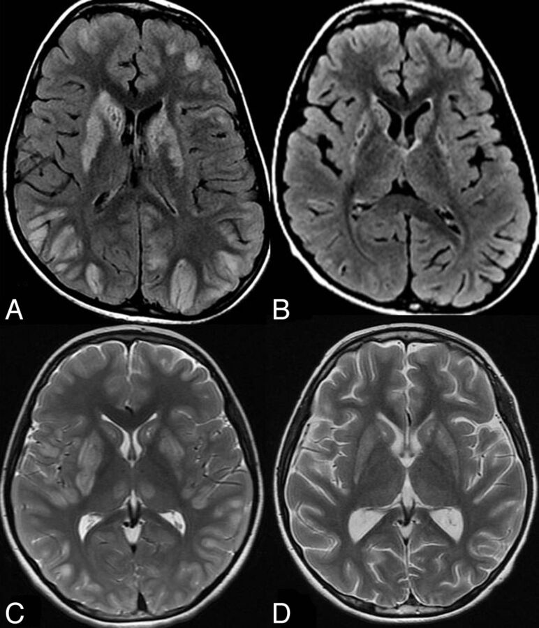
Fig 3.
Brain MR imaging in 2 different patients with BBGD (A and B, axial FLAIR for patient 1; C and D, axial T2WI for patient 2). In the 2 patients, the pretreatment images (A, C) demonstrate bilateral swelling and increased signal in the caudate nuclei and putamina with abnormal signal in the cortex and gray-white matter junction at multiple locations. Involvement of the medial thalami is also noted in patient 2 (C). Follow-up after treatment (B, D) shows disappearance of the abnormal signal in the cortical and subcortical lesions with resolution of the thalamic lesions in patient 2, who developed mild brain atrophy. The basal ganglia became atrophic with persistent abnormal signal intensity.

