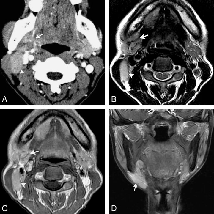
Fig 2.
LEC of the right submandibular gland in a 53-year-old woman with a painless mass in the right neck for 1 year. Axial contrast-enhanced CT (A) shows an ill-defined mass in the right submandibular gland with heterogeneous moderate enhancement (arrow). An enlarged lymph node in level IIa is also noted and shows a homogeneous enhancement (arrowhead). Axial T2-weighted image (B) shows a heterogeneous slight hyperintensity mass with curvilinear and linear hypointensity within it (arrow) and an enlarged lymph node in level IIa (arrowhead). Gadolinium-enhanced axial T1-weighted image (C) and coronal T1-weighted image with fat saturation (D) show the mass with a heterogeneous enhancement with hypoenhanced strands within it. An enlarged region of the lymph node in level IIa is also noted and shows homogeneous enhancement (arrowhead).

