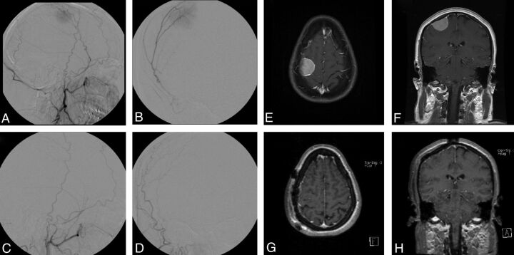
Fig 1.
Right frontal meningioma, pre- and postembolization and resection. Pre-embolization right ECA injection demonstrates tumor blush from the middle meningeal artery (A, lateral view; B, frontal view). Embolization was achieved with 150- to 250-μm PVA particles. Postembolization right ECA injection reveals complete obliteration of tumor blush (C, lateral view; D, frontal view). T1-weighted MR imaging with gadolinium before embolization (E, axial view; F, coronal view). T1-weighted MR imaging with gadolinium postresection (G, axial view; H, coronal view).

