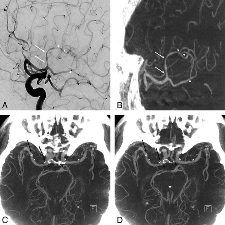
Fig 2.
An 81-year-old woman who underwent coil embolization of an unruptured posterior communicating artery aneurysm 1 month before was referred due to a history of progressive left hemiparesis. A and B, Good concordance was obtained for depicting stenosis at the proximal (arrows) and distal (arrowheads) M2 segment between DSA and IV FDCT. C and D, Axial maximum-intensity-projection image of IV FDCT allows a clear delineation of the vascular lumen from circumferential calcification (arrows) without superimposition of bone (arrowheads).

