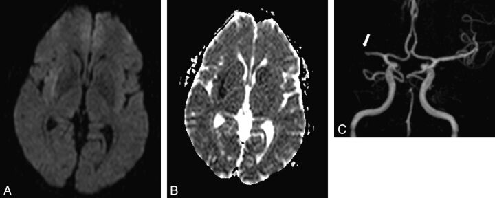
Fig 2.
A 59-year-old man with left regressive hemiparesis (clinical duration = 3 minutes, delay since the first clinical symptom = 3 hours). ABCD2 score = 4. A, DWI demonstrates a subtle hyperintensity in the right subinsular region with corresponding decrease of ADC on the ADC map (B). C, 3D TOF depicts right M1 segment grade 2 occlusion (arrow), responsible for the acute ischemic lesion in the insular lobe. The patient had another left transient hemiparesis in the subsequent 48 hours.

