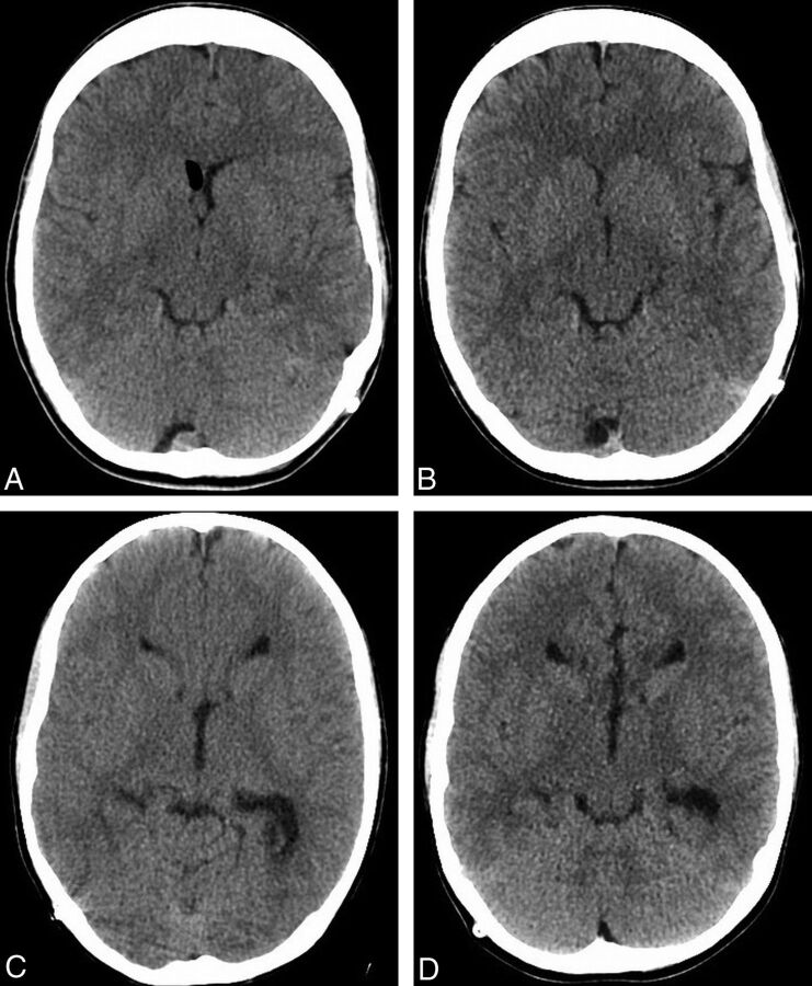
Fig 4.
Examples of study patients. Images from FBP (A) and ASIR (B) examinations performed 26 days apart in a 14-year-old patient, with a 23.7% decrease in the CTDI (34.9–27.5 mGy) and a 39.6% decrease in the DLP (581.5–389.3 mGy-cm) in the latter examination. Images from FBP (C) and ASIR (D) examinations performed 77 days apart in a 10-year-old patient, with a 26.4% decrease in the CTDI (32.3 to 24.7 mGy) and a 25.8% decrease in the DLP (452.3 to 349.0 mGy-cm) in the latter examination.

