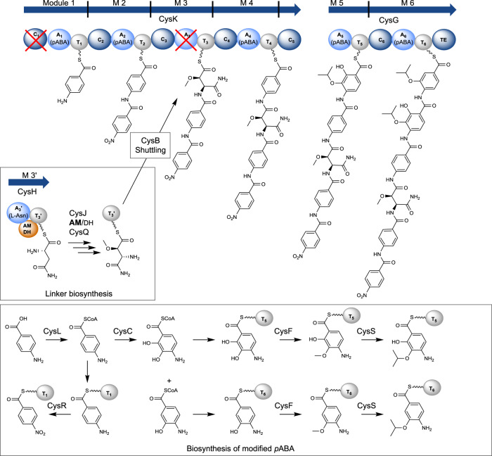
Fig. 7. Revised biosynthesis of cystobactamids on the example of Cys919-1.
C: condensation domain (dark blue sphere), A: adenylation domain (light blue sphere), T: thiolation domain (gray sphere), TE: thioesterase domain (dark blue sphere), AMDH: aminomutase dehydratase domain (orange sphere), red cross: inactive domains. Biosynthesis of the α-methoxy-l-isoasparagine linker moiety is described in more detail in Fig. 5. The 6-modular assembly line is encoded by cysK and cysG (blue arrows). CysR converts pABA to pNBA in trans or after product release from the assembly line. Biosynthesis of 2-isopropoxyl-pABA and 2-isopropoxyl-3-hydroxy-pABA is presumably catalyzed by CysC, CysF, and CysS. pABA is incorporated by M 1 and M 2, respectively. The linker moiety is transferred from T3’ (CysH) to T3 (CysK) by CysB (see Fig. 6). Another pABA is incorporated by M 4. Two tailored pABAs are incorporated by M 5 and M 6.

