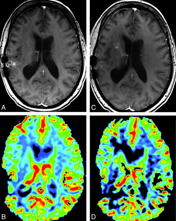
Fig 1.

A and C, Postcontrast SPGR image for reference MRI (C) shows new enhancement along the right anterior limb of the internal capsule, which was not present on comparison postcontrast SPGR (A). B and D, This area shows increased CBV on the concurrent DSC color map (D) compared with the prior DSC color map (B). The appearance was most suggestive of tumor progression, and the management plan was altered accordingly. Susceptibility artifacts are seen along the previous resection cavity in the right temporoparietal region on all images.
