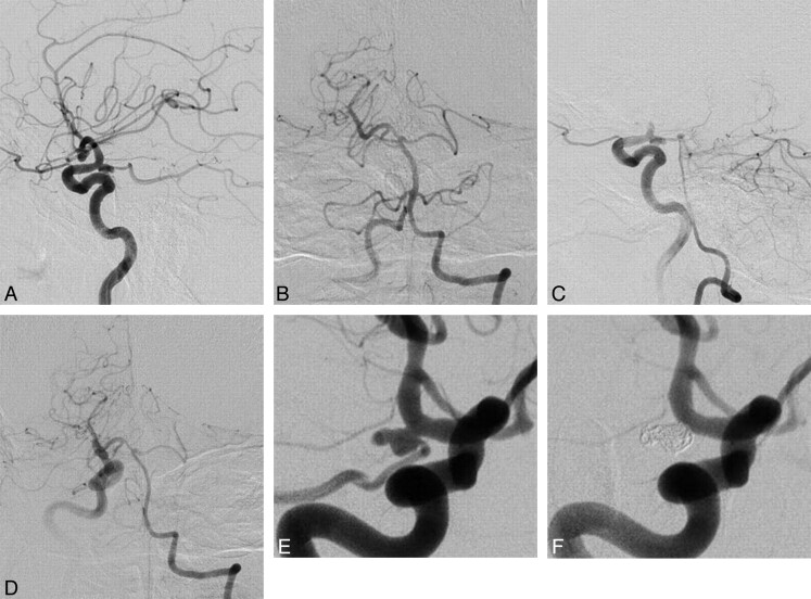
Fig 2.
A 69-year-old man without postoperative tuberothalamic infarction. A, Lateral view of the preoperative right internal carotid angiogram, indicating a posteriorly projecting right PcomA aneurysm with a maximum diameter of 6.9 mm. B, Anteroposterior view of the left vertebral angiography, demonstrating anterograde visualization of the right posterior cerebral artery (positive P1). C (lateral view) and D (anteroposterior view), The Allcock test revealed retrograde filling of the right PcomA and the aneurysm through the right P1. E, Preoperative right internal carotid angiography in a working projection depicted the right PcomA aneurysm with a PcomA arising from the aneurysmal sac. F, Right internal carotid angiography obtained immediately after coiling revealing the complete elimination of the aneurysm, along with the origin of the PcomA.

