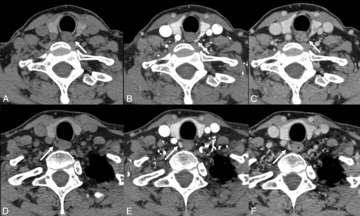
Fig 1.
A 63-year-old woman with primary hyperparathyroidism. No lesions were identified on sonography or technetium Tc99m sestamibi. 4D-CT demonstrates avidly enhancing lesions in the orthotopic superior location (arrows) bilaterally with rapid washout of contrast greater than that of the adjacent thyroid gland (A and D: noncontrast phase; B and E: initial postcontrast “arterial” phase; C and F: delayed postcontrast phase). This patient underwent bilateral exploration, and bilateral superior parathyroid adenomas were found at surgery.

