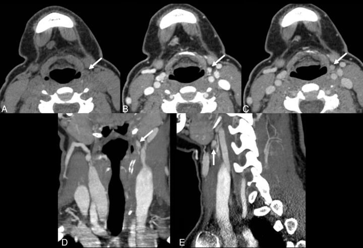
Fig 2.
A 26-year-old woman with persistent primary hyperparathyroidism after undergoing a 7-hour neck exploratory procedure including the upper mediastinum and a left hemithyroidectomy, as the left inferior gland could not be found. 4D-CT demonstrates a small lesion high in the left neck at the level of the hyoid bone (arrows). Perfusion characteristics are suggestive of a parathyroid adenoma, with a lesion lower in attenuation than the thyroid gland on the initial noncontrast phase (A), and rapid uptake of contrast (B) and rapid washout of contrast (C), greater than that of the thyroid gland (not shown). (D: coronal reformatted image in the “arterial” phase; E: sagittal reformatted image in the “arterial” phase). At surgery, a parathyroid adenoma was found in the left carotid sheath, at the apex of ectopic thymic tissue, consistent with an undescended left inferior parathyroid gland.

