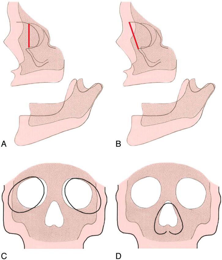
Fig 12.

Lateral diagram of the fetal skull (A) (darker areas) and the adult skull (B) (lighter areas) shows that the inferior orbital rim and the superior orbital rim are in the same plane in the young face (dark line in A), but in the older face, the supraorbital region protrudes forward of the cheek (dark line in B). Frontal diagrams (C and D) show that as the maxillary bones and the ethmoid bodies separate from one another, this movement increases the lateral growth of the interocular distance. As a result, the orbits enlarge and shift laterally (C). The movement of the nasal area combined with the malar movement results in an increase in the vertical size and the width of the upper part of each nasal cavity (D). (Modified with permission from Enlow D. A Morphogenetic Analysis of Facial Growth. Am J Orthodontics 1966;52:283–299. Figures 1 and 4)
