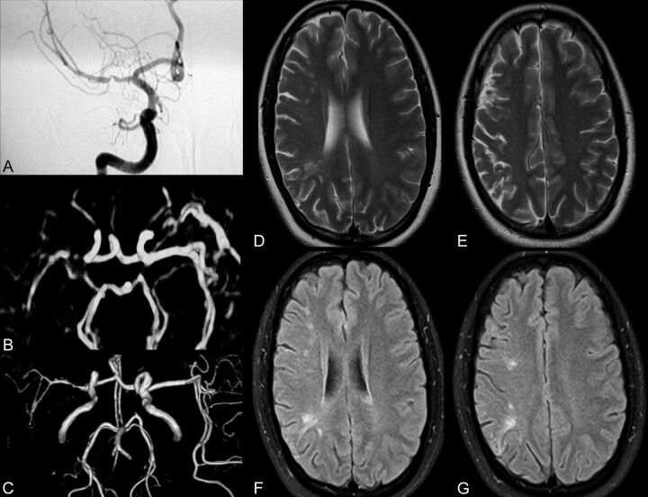
Fig 1.
Comprehensive imaging of a patient with recent stroke depicting left MCA stenosis. A−G, DSA (A) confirms contrast-enhanced MRA (B) and volume-reduced TOF MRA (C) findings of severe stenosis within the right MCA in a recently symptomatic patient with small ischemic lesions seen on T2 (D and E) and FLAIR (F and G). MRA (B and C) overestimates the degree of stenosis in this particular case.

