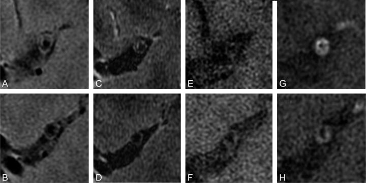
Fig 3.
Multispectral high-resolution MR imaging of a symptomatic patient with MCA disease. T2-weighted (A and B), spin-echo inversion recovery (C and D), T1-weighted (E and F), and postgadolinium T1-weighted (G and H) images depict a large eccentric contrast-enhancing plaque within the lumen of a recently symptomatic patient on the ipsilateral side of symptom presentation.

