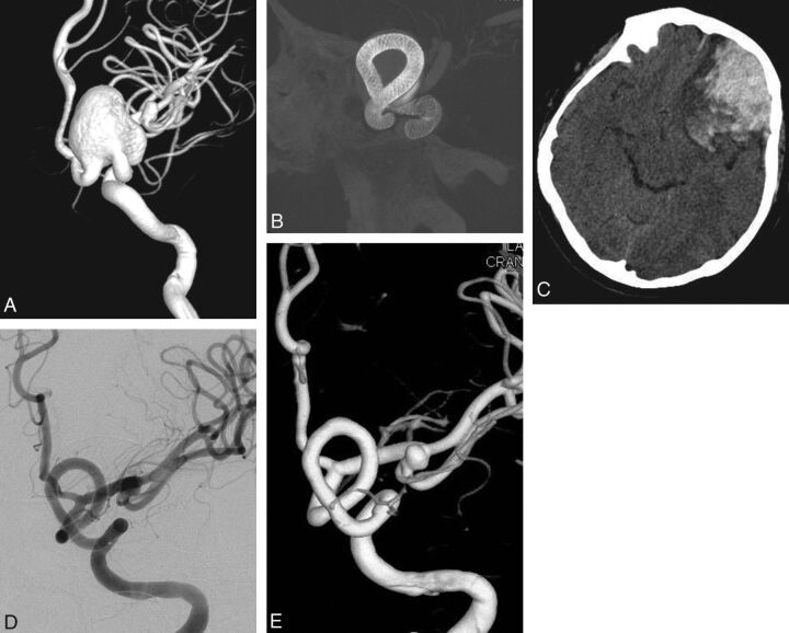
Fig 1.
Preoperative 3D angiogram (A) shows a very wide-neck large ICA aneurysm. It could be reconstructed with several overlapping devices, creating a new vessel wall within the sac as seen on the perioperative DynaCT image (B). Postoperative CT obtained the same evening (C) reveals ipsilateral frontal intraparenchymal hemorrhage. 2D (D) and 3D (E) views of 6-month control angiography demonstrate the reconstruction of the parent artery and total occlusion of the aneurysm.

