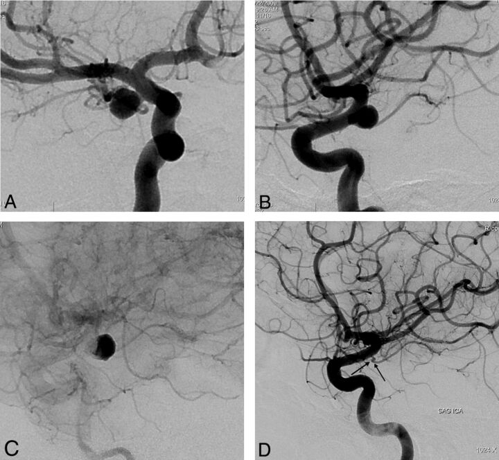
Fig 6.
Preoperative 2D (A and B) angiograms show the ICA aneurysm in which the anterior choroidal artery is originating from the aneurysm at the neck. A single PED is placed covering the neck, causing stagnation of the contrast within the sac (C). Six-month control angiography (D) demonstrates total occlusion of the aneurysm with the anterior choroidal artery preserved (arrow).

